


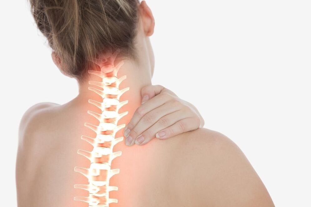
The spine, like any structure that performs a support function, inevitably wears out over time. High static and dynamic loads and local overloads of the particularly mobile segments of the upper part lead to a decrease in regenerative capacities and a progressive degeneration of the neighboring cartilaginous and musculo-ligamentous structures. By the age of 30-35, almost everyone has more or less significant signs of cervical osteochondrosis. And if it is impossible to stop the irreversible process of biological aging, it is quite possible to slow it down.
Diagnostic
For objective assessment of the condition and detection of degenerative-dystrophic changes in the cervical spine, X-ray imaging methods are used:
- simple spondylography (radiographic study without contrast in frontal, lateral and oblique projections)
- x-ray with functional tests
- MSCT (multi-slice computed tomography)
- MRI
- Spondylography by probing of the upper spine is a traditional method of radiological diagnosis of cervical osteochondrosis. With its help, the condition of the vertebral bodies is assessed, their shape, height, degree of deformation and displacement with respect to each other are determined. On x-ray images, osteophytes, areas of illumination in foci of liquefaction of bone tissue are visualized.
- Spondylography with functional tests is a study to identify signs of movement disorders. The x-ray is performed with a fixed maximum flexion and extension of the cervical spine.
- MSCT is a progressive alternative to X-rays. Bone structures, intervertebral discs, ligamentous apparatus, spinal canal and spinal cord are visualized in more detail on multi-layered images.
- Magnetic resonance imaging allows additional visualization of the cartilage layer and other soft tissues of the spinal joints. The study is prescribed for severe neurological symptoms, to differentiate cervical osteochondrosis from acute intervertebral hernia.
Treatment of cervical osteochondrosis
Treatment of osteochondrosis of the cervical spine is aimed at eliminating pain and slowing the progression of the pathological process. It is carried out in two directions: to limit the impact of unfavorable factors and to suppress the mechanisms of development of the disease.
Therapeutic and prophylactic measures that minimize the impact of the causative agents include:
- rational selection of work furniture
- use of orthopedic mattresses and pillows
- correction of hearing, vision and posture
- wear special fasteners
- restriction of work activities associated with a long stay in a forced situation
- adequate physical activity
- adequate nutrition
There are many methods of therapeutic correction that are designed to slow the development of the degenerative process.
Massage for cervical osteochondrosis
Massage procedures aimed at relieving inflammation and eliminating pain are included in the set of mandatory therapeutic measures. The most effective types of neck massage:
- classic
- medical (manual)
- tip (acupuncture)
- vacuum packed (canned)
- Equipment
Thanks to massage techniques, local blood and lymphatic circulation is improved, tissue trophism is accelerated, muscle pinching is eliminated, neck tensions are relieved, muscle tone and elasticity are improved.
Orthopedic collars
To fix the cervical spine in the correct position, special orthopedic devices (Shans collars) are used. Removable structures of various sizes, shapes and degrees of rigidity limit the usual pathological position of the head, control the movements of the neck, reduce pressure on the vertebral segments, warm and relax tense muscles and prevent the progression of the disease.
The cervical collar for osteochondrosis is available in several versions:
Soft medical foam splintsor other porous hypoallergenic materials have a notch for the chin and lower surfaces of the neck, and retainers. They are used to correct minor disorders of the upper spine, to keep the vertebrae in an anatomically correct position, and to relax the muscles of the shoulder girdle.
Pneumatic collars (inflatable)are intended for pain prevention, gentle traction and the elimination of compression of the vertebral artery.
Semi-rigid dressingsequipped with metal inserts reliably stabilize the intervertebral segments. They considerably limit the range of motion and contribute to the widening of the spaces between the vertebral bodies.
Durable plastic rigid corsetsdesigned to completely immobilize the cervical spine in a neutral position. Prescribed in advanced stages of the disease, accompanied by compression disorders.
The collar for osteochondrosis of the cervical spine is chosen by the doctor taking into account the age, anatomical features and stage of the degenerative process.
Manual therapy
Manual therapy aims to identify and remove blockages in the motor segments. A local dosed effect on the spinal joints helps normalize blood flow and blood supply to the brain, eliminate compression (pinching) and restore normal functioning of nerve fibers. Specific manipulations by the chiropractor allow you to achieve maximum relaxation, eliminate muscle spasms, cervicogenic headaches resulting from damage to the anatomical structures of the neck and tension headaches.
Acupuncture
Acupuncture, involving the installation of acupuncture needles in bioactive points of the neck and shoulder blades, aims to restore the disturbed energy balance. By stimulating rapid contractions of sensitive nerve fibers and the release of endorphins and neurotransmitters, acupuncture for cervical osteochondrosis has a powerful anti-inflammatory and analgesic effect. Thanks to this technique, numbness of the hands, dizziness, tinnitus, improves blood circulation and optimizes mobility.
Physiotherapy
Physiotherapy of degenerative pathologies of the spine is aimed at relieving pain and stimulating recovery processes. The greatest therapeutic effect is provided by:
- UFO
- ultrasound treatment
Faq
How to provide assistance during acute pain with osteochondrosis of the lumbar spine?
With sudden sharp pain, it is necessary to fix the lower back. This will immobilize the spasmodic muscles and shift the load off them. Then, if possible, lie the patient on their back with a pillow placed under the bent knees. To reduce the pain, you need to take medication with analgesic and anti-inflammatory effects (NSAIDs). In addition, you can use an ointment or gel based on diclofenac or its analogues, or apply a cold compress (no more than 10 minutes). It is very important to eliminate stress on the spine and to consult a doctor as soon as possible.
Is it possible to do physical exercises for lumbar osteochondrosis?
Physical education for lumbar osteochondrosis is not only prohibited, but also recommended (except for a period of acute pain). However, you should be careful not to allow an axial load on the spine and categorically refuse to squat, jump and lift weights. The set of exercises should be selected by a specialist on an individual basis.